

Why Before-and-After Images Don’t Tell the Whole Story
Define the frame first: a before-and-after set is a protocol, not a promise. In procedures with forehead wrinkle filler, what you see depends on how the image was taken as much as what was done. Clinics post forehead wrinkle filler before and after galleries to guide you, yet they often mix time points, lighting, and angles. Data matters. Hyaluronic acid gels typically persist 6–12 months, swelling peaks at 24–72 hours, and muscle tone varies across the day. If frontal is activated in the “before” shot and relaxed in the “after,” lines fade even without change — funny how that works, right?

What do before-and-after photos prove?
They show a moment, not the mechanism. The hidden pain points are subtle: head tilt, brow raise, skin hydration, and even makeup blur fine lines. Product science adds another layer. Cross-linking density changes viscoelasticity and G-prime (G’), so two syringes can flow and lift very differently. A high G’ gel placed on the supra-periosteal plane supports the skin well, but a superficial pass may show sheen or stiffness. Traditional photo-only reviews miss these variables. Look, it’s simpler than you think: ask how the images were standardized and how the rheology matched the crease depth. That way, you read the gallery as data, not drama. Next, let’s move from pictures to principles.

From Gallery Glare to Guided Precision
What’s Next
Forward-looking clinics now use new technology principles to make results repeatable. Ultrasound mapping charts vessels before injection, so the cannula path avoids hotspots and reduces bruising. 3D imaging captures volume change under identical lighting, letting you compare like with like. Even better, AI tools can normalize white balance and head position, so progress is not a pose. Pair this with rheology-aware selection of dermal fillers for forehead wrinkles: choose cohesivity for contour hold, diffusion kinetics for soft blending, and a G’ that matches the crease load. The outcome? Smoother arcs without a waxy look — the kind of “after” that still moves.
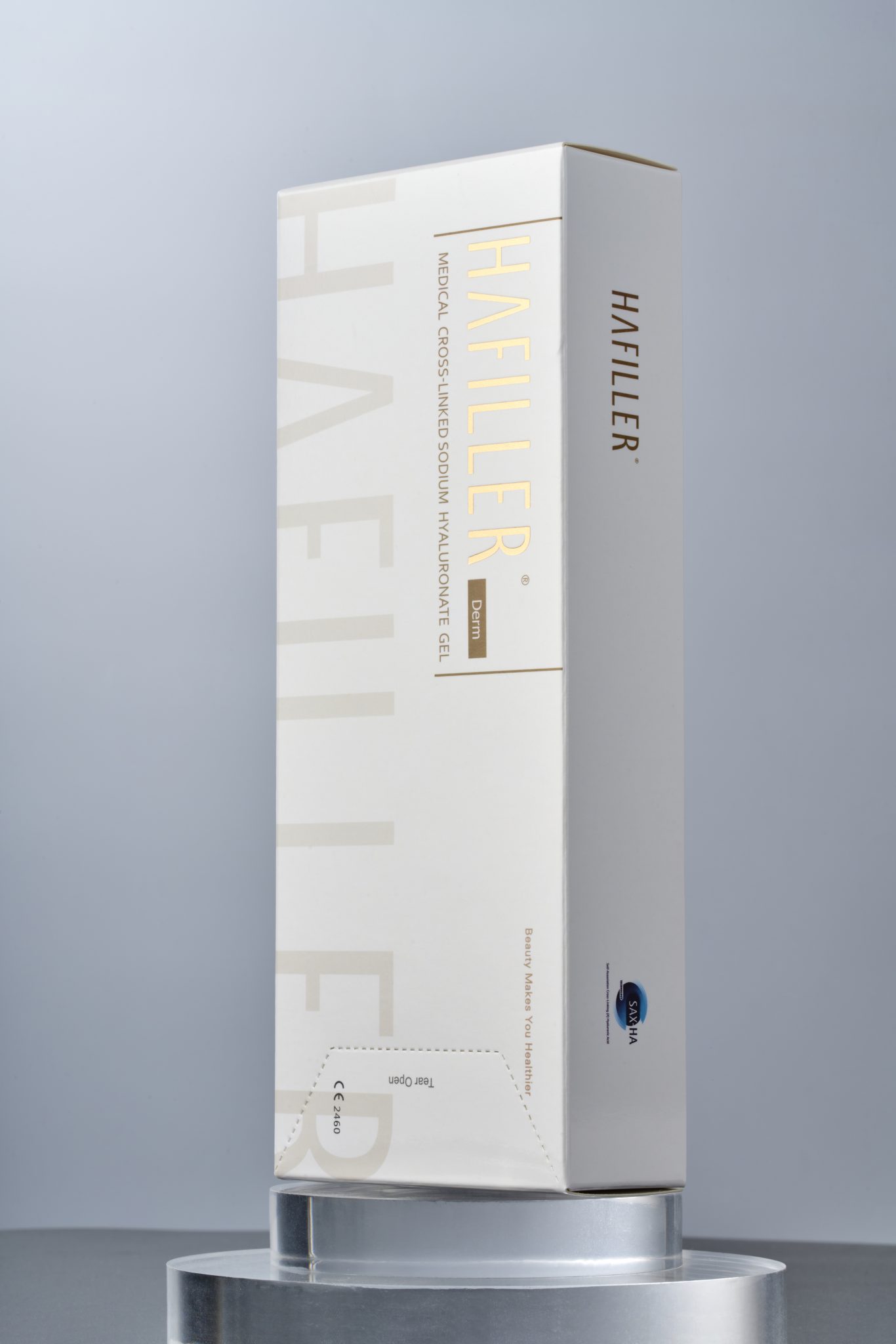
So where does that leave you? Think comparative, not absolute. Photos are a start, yet protocols are the proof. Summarizing the signal: images need standardization; products need property fit; techniques need controlled depth. For an advisory close, use three metrics when you choose a provider or plan: first, evidence of standardized imaging (same lens, light, time post-procedure); second, documented product rationale linking cross-linking density and G’ to your wrinkle type; third, technique transparency, including plane of placement and cannula vs needle choice. Small checks, big clarity — and fewer surprises on your own “after.” For further technical reading, see HAFILLER.